![]()
Catherine ForseM.Sc Candidate; Department of Molecular & Medical Genetics
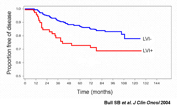
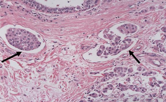
Identification and characterization of genes involved in lymphovascular invasion in axillary node-negative breast cancerAn important prognostic variable that dictates the progression of early breast cancer is the number of involved axillary lymph nodes. Approximately 20% of women with axillary node-negative (ANN) breast cancer will experience a recurrence. Currently, there is no single predictor of outcome for ANN breast cancer; however, prognostic markers have been described. Our lab has identified the presence of lymphovascular invasion as an alteration of ANN breast tumours that can be used to predict patients likely to experience a recurrence [1]. Lymphatic invasion (LVI) is defined as the presence of tumour tissue within peritumoral lymphatics, capillaries or postcapillary venules. Although LVI is associated with poor outcome in ANN patients, very little is known about the cellular alterations associated with the presence of LVI in breast tumours. The objectives of my project are (i) to identify genes which have altered expression in tumours with and without LVI in ANN breast cancer patients and (ii) to investigate the mechanism by which a gene and/or pathway of interest may promote cancer progression using in vitro assays. FIGURES: 1 and 2 Lymphovascular Invasion (LVI) is an independent prognostic factor for ANN breast cancer
|


.")

