![]()

Cardiovascular and Placental Physiology During Pregnancy
|
||
|
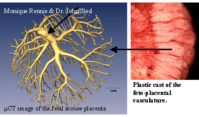
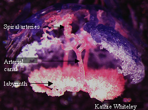
Our lab has a longstanding interest in cardiovascular hemodynamic development in general, and in the role of the placenta in controlling maternal and fetal hemodynamic function during pregnancy in particular. Our current work investigates the feasibility and tremendous potential of performing physiological measurements in genetically-altered mice to explore the mechanisms involved. Using large animals and computer models, we showed that elevated vascular resistance in the feto-placental microvasculature and/or venous outflow tract most likely causes the highly pulsatile blood velocity waveforms commonly observed in the umbilical arteries of human fetuses with intrauterine growth restriction. We are now using high frequency ultrasound to monitor umbilical arterial velocity waveforms in mouse embryos, and vascular casting techniques and micro-computed tomography to evaluate placental vascularization during pregnancy in normal and mutant mice. Similar methods are being used to simultaneously evaluate cardiovascular function in the maternal circulation during pregnancy. The goal of this work is to determine the developmental mechanisms responsible for abnormal placental hemodynamics and the impact on fetal growth and development, and on maternal cardiovascular adaptations to pregnancy. Ultimately, this work will advance our understanding of two of the most common and serious complications of human pregnancy, fetal intrauterine growth restriction and maternal preeclampsia.
|
||
|
Doppler blood velocity recorded from the umbilical artery of a mouse embryo at 15.5 days of gestation. Click on the speaker to hear how it sounds. |
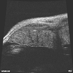
UBM movie by Dr. Dawei Qu Using an Ultrasound Biomicroscope, the placenta can be imaged through the mother’s skin (she is anesthetized during the procedure). Fetal blood can be seen flowing in the blood vessels in the placental labyrinth in this movie. |
|


.")

