Lunenfeld-Tanenbaum Research Institute

BIOMINERALIZATION

Changes in the quality of bone with age and disease


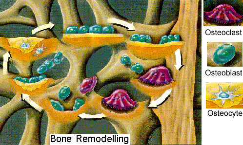
Mineralization profiles are a much more sensitive index of tissue aging than chronological age. In normal physiological conditions, there are shifts in mineralization towards lower densities when turnover is high and towards higher densities with aging. We are studying the correlation between these changes and the lowering of bone mechanical properties which results in fractures. In the case of postmenopausal osteoporosis (high turnover), where we have found a shift to lower mineralization. We are investigating the changes in cancellous bone architecture and material properties as the mechanism leading to vertebral fractures. In the case of age-related osteoporosis, we have shown that there is a non linear increase in mineralization with age in humans together with a decrease in macroscopic density and an increase in porosity. It has also been shown that microcracks accumulate with age in cortical bone. We are therefore investigating the hypothesis that increased mineralization leads to accumulation of microcracks leading to fatigue damages and to cortical bone fractures.

bone remodeling

Both major functions of the skeletal system (ion homeostasis and mechanical support) are dependent on the chemical nature, size, shape and orientation of the mineral component. It is also dependent on the interaction between the mineral and the organic matrix because soluble, mineral bound and collagen bound proteins have different effects on the fabric of bone. Because of the rates at which calcified tissues are turned over, there are populations of mineral particles of different ages and properties in every sample. Therefore, the changes in chemical and structural characteristics of the mineral component and its interaction with the organic matrix during formation, maturation and resorption need to be understood to interpret changes in the quality of the bone fabric at a molecular and tissue level to distinguish between normal and pathological bone loss.

Pathological mineralization
In collaboration with a group in the USA, who has developed the first rat model of kidney stone, we are looking at the mechanisms of their formation and at the regulation of pH balance in the body by the bone mineral carbonate from the skeleton. We are trying to relate the information from the animal model to human stone formation to devise strategies to prevent their formation.

The structure and chemistry of bone mineral and matrix
We are interested in the basic structure and chemistry of mineralized tissue, with a particular emphasis on bone. As bone mineral changes with age, location and rate of turnover, we have to distinguish between local areas which have just mineralized and areas which contain mature bone crystals. By establishing mineralization profiles which separate newly formed bone from progressively maturing bone, we can analyze each fraction separately to determine crystal size, mineral chemistry and crystal packing in the bone matrix. By studying the mineral matrix interaction of the extracellular matrix of bone, we are trying to understand the mechanisms of mineral deposition, maturation and resorption. We have also analyzed the changes in the distribution of soluble, mineral bound and collagen bound proteins of bone with age and in osteoporosis to understand the relation between the mineral and the organic matrix of calcified tissues. We have shown that bone mineral does not contain amorphous calcium phosphate even at the earliest embryonic stage of mineralization but was made of poorly crystalline apatite with many lattice substitutions and a changing composition with age and maturation. We are in the process of following bone mineral crystals in the chick from the beginning of mineralization (8 days embryo) to old age (70 weeks old chicken), using x-ray diffraction, neutron activation analysis and infra-red spectroscopy.

Future Research
The effect of collagen defects on the mineralization of bone in osteogenesis imperfecta.