![]()
Elemental Chemical AnalysisOur laboratory analyzes for bone mineral content using various methods. Calcium(Ca) and Phosphorus(P) Assays are performed to using a colourimetric reaction to determine ion content. The intensity of the colour reaction is directly proportional to the content of Ca and P in the bone sample. Carbon Detection is performed by using a carbon coulometric system which measures carbon as CO2. Trace Eements found in bone, such as fluoride or strontium are quantified using a Slowpoke nuclear reactor or by exciting protons and measuring their decay by Backscatter Electron Microscopy (BSE).
CALCIUM & PHOSPHATE ANALYSISPhosphate Assay In an acid medium, molybdate is transformed into a heteropolyacid, which subsequently reacts with phosphate to produce a complex. Molybdate exists as a dimer, which polymerizes and reacts with phosphate to form duodecamolybdophosphoric acid (12 MPA). 12MoO4 + H3PO4 + 24H ---> H3PMo12O40 + 12H2O Upon reduction, the acid forms a blue product, referred to as "heteropoly blue" This colourimetric method is based on the method of Chen, P.S., Toribora, T.Y., Warner, H. (1956; Microdetermination of phosphorus. Anal Chem. 28:1756-1760). The ashed sample is mixed with an acid solution of ammonium molybdate which forms phosphomolybdic acid by combining with the phosphate present. The acid is reduced by the the addition of ascorbic acid to produce a blue colour whose intensity is proportional to the amount of phosphate present. Calcium Assay In this method, a cresolphthalein dye is used to form a complex with Ca2+, which is violet coloured at alkaline pH. This colourimetric method is based on the principles described in Schwarzenbach, G. (1955; The complexones and their analytical application. Analyst 80, 713 - 729). The ashed sample is mixed with an alkaline solution of cresolphthalein complexone (CPC), a.k.a. Phthalein Purple, buffered with sodium borate which forms a o-CPC complex by combining with the calcium ions present. The colour intensity of the complex formed is directly proportional to the calcium concentration. Ca2+ o-CPC --- alkaline pH ---> calcium-o-CPC complex
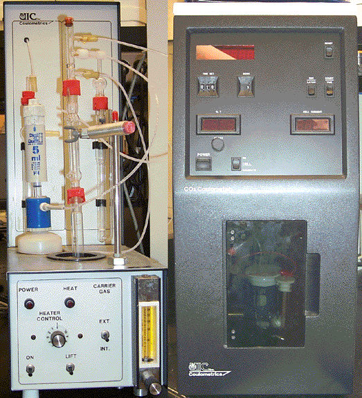
CARBONATE ANALYSIS
Principles The acidification module acidifies bone samples to evolve inorganic carbon as CO2. The potassium hydroxide (KOH) scrubber removes CO2 from air. Air free of CO2 sweeps the evolved CO2 into the coulometer. The coulometer determines carbon content in any CO2-containing gas stream. The coulometer cell contains a solution of monoethanolamine (MEA) and a pH indicator, a platinum cathode and a silver anode. The assembly is positioned between a light source and a photodetector in the coulometer. The CO2 in the gas stream from the acidification module enters the cell. It reacts with the MEA to form a titratable acid, indicated by a fading of colour of the coulometric solution: CO2 + HOCH2CH2NH2 -----> HOCH2CH2NHCOOH Photodetection records the change solution colour as percent transmittance (%T). As %T increases, the titration current electrochemically generates base, with e- generated at the silver anode. The reactions are: 2H2O + 2e - ----> H2(g) + 2OH - Ago ----> Ag+ + e - The silver anode is gradually used up. The silver ion reacts with excess potassium iodide. Neutralization occurs as:
INAA ANALYSISTrace elements in bone may be determined by Instrumental Neutron Activation Analysis (INAA). INAA may be defined as a chemical element analysis made by nuclear activation followed by the measurement of specific induced radioactivities without the use of radiochemical separations. This type of analysis is performed on a SLOWPOKE (Safe LOW POwer Kritical Experiment) Reactor with a gamma-ray counting detector. The SLOWPOKE Facility can analyze samples for a number of elements simultaneously, with a sensitivity at the ppm level. The analytical techniques used require minimal sample preparation and are non-destructive permitting retention of valuable samples or re-use of the same sample for further measurements. For our studies, we are primarily interested in the following bone elements:
BACKSCATTERINGPrinciples In an electron microscope, electrons are released from a an electron gun which are then accelerated by a potential difference and focused by magnetic condenser lenses onto the (bone) sample. An electron passing close to a nucleus in the sample will be deflected by the sample's electric field. When an electron is deflected by more than 90o, it is said to be "backscattered". The deflected electrons will be backscattered onto a back-scatter electron (BSE) detector which is positioned directly above the sample. The probability of an electron being backscattered depends on the nuclear charge and the inter-nuclear distances. The degree of mineralization affects the number of nuclei available to backscatter electrons.
|

.")

