![]()
Histomorphometry
Histomorphometry is broadly defined as the measurement of the shape or form of a tissue. Quantitative analysis of bone architecture is achieved using bone histomorphometry which provides valuable information on the amount of bone and its cellular activity. Structural parameters evaluating the quantity of cancellous bone and osteoid are measured using static histomorphometry. To study the changes in cellular activity over time, the bone is double labeled with a fluorochrome and measured using dynamic histomorphometry. Together, the information obtained from both static and dynamic histomorphometry provides a useful profile of bone turnover.
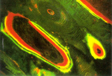
Dynamic Histomorphometry We employ the tetracycline double labeling technique for the histomorphometric evaluation of bone dynamics. The uptake of tetracycline by the bone allows us to evaluate the status of mineralization over a period of time (usually 10-14 days). The figure above (from Bone Histomorphometry, Eriksen et al., 1994) demonstrates a calcified bone sample which is stained with Goldner’s Trichrome. Mineralized bone is stained green and osteiod is stained red. Similar to static histomorphometry, the calcified bone samples are processed and embedding in plastic resin. However, dynamic histomorphometry is conducted on unstained bone sections and the tetracycline double labels are measured under fluorescent light. The dynamic parameters of bone turnover include mineral apposition rate, mineral formation rate and mineralization lag time. This will ultimately provide us with a quantitative assessment of the extent of bone formation over a specific period of time.
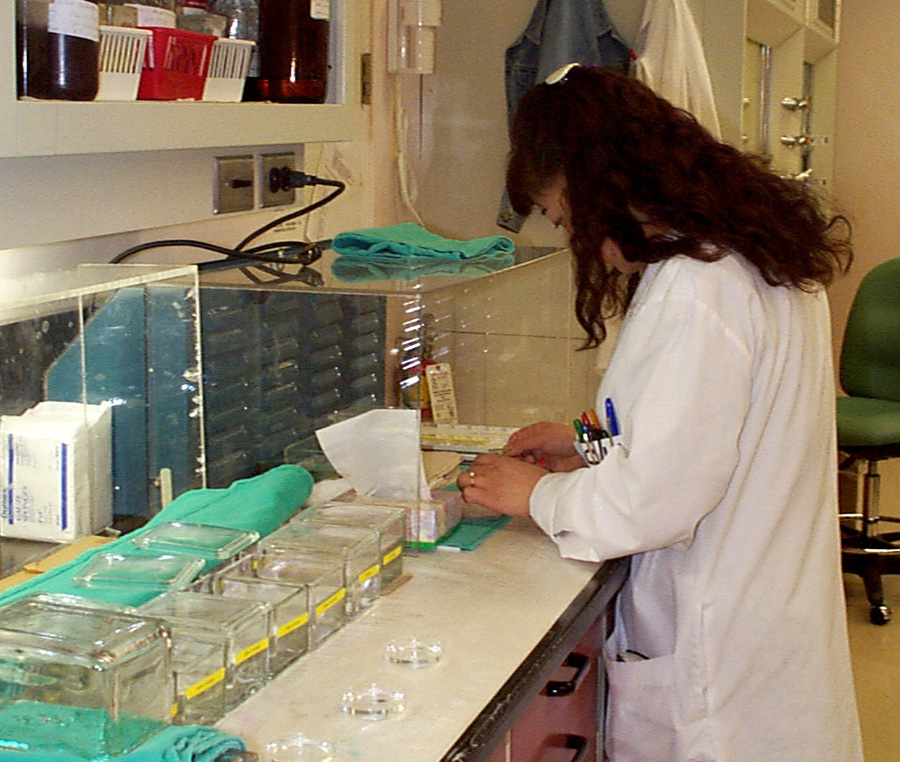
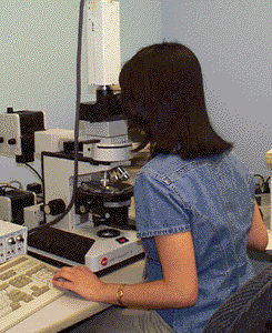
Static Histomorphometry Static histomorphometry allows us to quantitatively evaluate the structure of bone at a particular time point (as opposed to dynamic histomorphometry which allows us to study bone over a period of time). Static indices of bone architecture include parameters such as the volume, thickness and separation of trabecular bone. Measurements of osteoid surface and volume and are also quantified to gage the extent of bone formation and bone resorption at the time of sample collection. In our laboratory, the calcified bone samples are processed and embedded in a plastic resin. The bone sections are subsequently stained with Goldner’s Trichrome to differentiate calcified bone from osteoid. Histological structures of the bone sample are manually measured by tracing the area of trabecular bone and osteoid using a digitizing tablet and a light microscope (right figure). The measurements of all the histological structures of interest are then obtained using computerized image analysis (Bioquant, R&M Biometrics). |

.")

