![]()
X-ray AnalysisBone structure may be examined using several techniques employed in this laboratory
IMAGE ANALYSISPrinciple: Mathematical Morphology is the application of lattice theory to the study of phenomena which spread in space and which exhibit a spatial structure. Developed in France by G.Matheron and J.Serra as a method for Image Analysis, the application of mathematical morphology is extended from the geological and mining field to metallurgy,remote sensing,robotics,medical sciences etc. Quantification of bone structure The images of bones (trabecular and cortical), containing structural information which is given by the totality of relationships among the pixels of the bone phases, are studied with a LEICA QUANTIMET 500IW Image Processing and Image Analysis System of Mount Sinai Hospital (Toronto, Canada). For trabecular bone analysis, connectivity and orientation measurements are taken in order to obtain : proportion of trabecular bone, average trabecular thickness, trabecular number, trabecular separation, end points, multiple points, cortex points, different categories of struts, anisotropy and the star. QUIPS programs containing mathematical morphological transformations (erosion, dilation, opening, closing, skeletonising, rose of directions etc.) are developed. For cortical bone analysis, grey-tone images containing structural information about the osteons and Haversian canals are processed with QUIPS programs. In order to obtain the values of bone parameters (total cortical area, osteonal area, mean osteonal wall thickness, osteonal density, total porosity, area of haversian canals and mean canal area), image processing operations (filtering, histogram equalisation) and Mathematical Morphological transformations like erosion, dilation, opening, closing and size distributions of the openings of the osteonal space are performed.
DIFFRACTION
DEXA
Jergas, M.A., Breitenseher, M., Glüer, C., Yu, W., Genant, H.K., 1995; Estimates of volumetric bone density from projectional measurements improve the discriminatory capability of dual x-ray absorptiometry, Journal of Bone Mineral Research, 10(7):1101-1110. For our purposes, BMD and BMC measurements of research animals provide useful information of observable trends in different test groups.
PIXIMUS
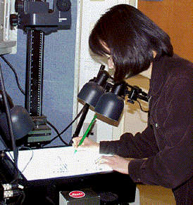
FAXITRON
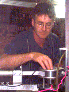
Faxitrons are used to preserve an image of bone samples which must be measured in a particular plane (ie. Femoral head fracture analysis) or to demonstrate areas of dense bone formation in a sample.
|
 Bone mineral structure can be analyzed by means of a X-ray diffractometer. Crystal (hydroxyapatite) size is determined by measuring the cross-section (D-130) and length (D-002).
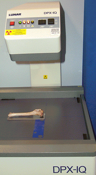 Dual Energy X-ray Absorptiometry (DEXA) is a common tool used by clinicians to evaluate fracture risk amongst patients. DEXA measures bone mineral density (BMD), which is a measurement of the bone mineral content (BMC) per unit area (this is due to the fact that DEXA is a projectional technique). BMD has been strongly correlated to bone strength and fracture risk. Since the third dimension of bone is not accounted for, it is not a true volumetric density and therefore there is a dependancy on size (a larger bone with the same volumetric BMC would yield a larger BMD than a smaller bone) (Jergas, et al. 1995).
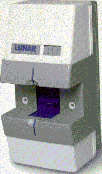 The PIXImus is a densitometer designed to specifically measure bone mineral density and body composition from total body imaging of a mouse. This smaller version of the larger DEXA (dual-energy x-ray absorptiometry) machine allows for multiple measurements of the mouse in situ over the life of the animal.

.")

