OptIma at Lunenfeld-Tanenbaum Research Institute
main Nikon/Upright SpinningDisc/Decon Leica Inverted/Dissecting Orde

OPTIMA, the Optical Imaging Facility
at Lunenfeld-Tanenbaum Research Institute

The OPTIMA Facility is a core technology operation that provides expert advice and maintains Microscopy and Imaging instruments. The systems are primarily used to image fluorochromes and histological stains, though bright field and reflected light images can be collected. Equipment range from microscopes equipped with cameras for fluorescence and transmitted light imaging to laser-scanning imaging systems. We train users to perform their own acquisition and analysis, yet also provide staff-assisted imaging services to internal and external scientists.

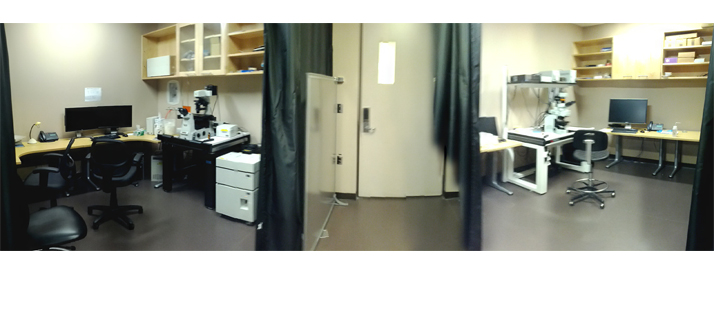
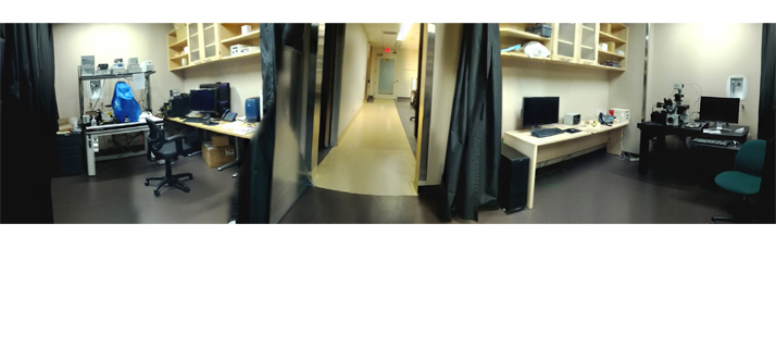
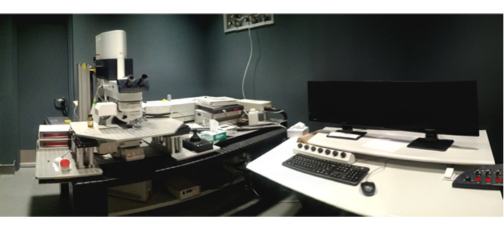
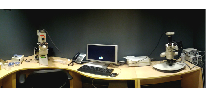
The Microscopy and Imaging Systems are located on the eighth floor Research Area and also on the sixth floor of the Orde/Murray Research Site. There are 2 confocal microscope systems (one is 2-photon), 2 spinning-disk confocals, a deconvolution-based Deltavision system, 2 Optigrid structured illumination fluorescence and histology camera systems, and multiple other basic camera-based imaging systems. In addition to classical microscope-based imaging systems, OPTIMA operates automated high-content screening systems (one spinning-disk).

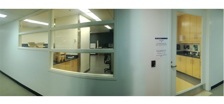
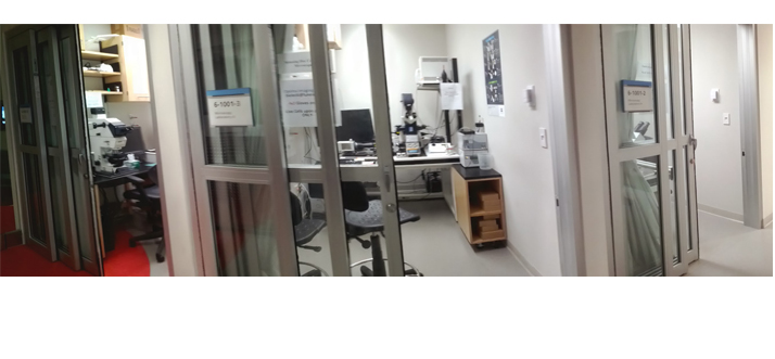
At the MSH eighth floor site, three staff members, Louise Brown, John Georgiou, and Sarang Kulkarni supervise the day-to-day operations. Four systems are housed in the 2010-PhaseI Imaging Suite located in Rm.871; the 2012-PhaseII expansion created our new main entrance via Rm.865, which houses an additional five microscope systems and four high-content screening imaging systems that are managed by Mikhail Bashkurov. The Microscopy and Imaging systems that are located at the Orde/Murray sixth floor Research Area are managed by Ryszard Bielecki. The Lunenfeld-Tanenbaum Research Operations division and the Imaging Committee, headed by Profs. Joe Culotti and Jim Dennis meet regularly to discuss various operational matters.
MSH Status:

  • Nikon Confocal
  • Leica Confocal
  • Deconvolution
  • Upright
  • Inverted
  • Dissecting
  • Stereomicroscope
Orde Status:

  • Quorum Spinning Disc
  • Optigrid Inverted
  • Olympus Stereology
  • Leica upright
  • Zeiss Axioplan upright
  • Leica DMIL LED inverted
  • Viral Tissue Culture
MicroscopeBooking:

ContactUs:

News

PrevNext

*New Stereomicroscope: The Nikon SMZ1500 captures images or video in real-time. Superior contrast - depth perception and visualization of green/red fluorochromes.


October 2013

New access: The imaging facility entrance is now via the eight floor central corridor, Rm 865. Contact us if your MSH card access is not working.


September 2013