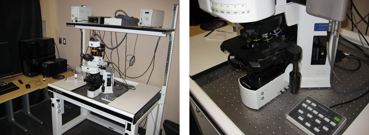
| Location | 865 (SW) |
| System Type | Optigrid-SIM/histo-fluor (camera-based) |
| Model | Olympus |
| Microscope | Olympus BX61 |
| Lenses | Dry 4,10,20/0.75 Oil 40x/1.3, 60x/1.42, 100x/1.4 |
| Vibration isolation | Yes |
| Stage | Manual xy, Slide spring clip |
| Z-focus | Integrated objective z-drive |
| Illumination | EXFO X-Cite 120
|
| Filtering | DAPI, FITC, TxRed, CFP, YFP, BF-brightfield |
| Detector | a) Fluor camera: Hamamatsu DP71 camera |
| | b) Color camera: Olympus DP71 |
| Pinhole | - |
| Software | Volocity / Xp |
| | DP Controller |
| Offline analysis | |
| Other features | QIOPTIQ Optigrid |
| | Structured illumination Microscopy (SIM) |
| | Deconvolution |


